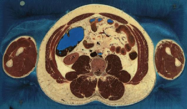
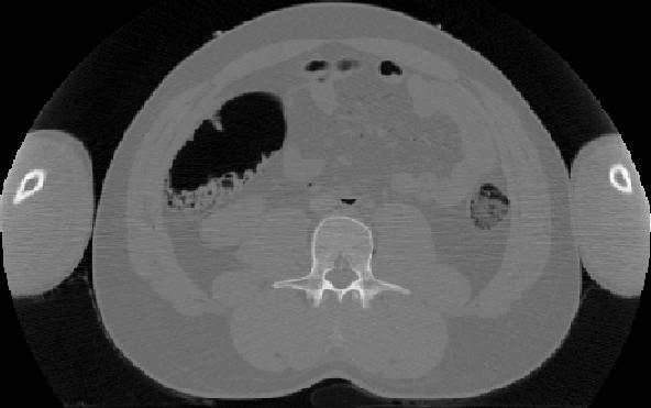
CT data for torso
The images shown on this page has been obtained from the Visible Human Project. They were created from scanning a human cadaver with CT and MRI scanners and then subsequently slicing the cadaver into 1 mm sections for taking photographs.
The CT data consists of axial CT scans of the entire body taken at 1 mm intervals at a resolution of 512 pixels by 512 pixels where each pixel is made up of 12 bits of grey tone.
A further description of the data can be found at here and a description of the program can be found here.
 |
 |
|---|---|
| Optical image | CT image |
Data for the CT-image can be obtained from the ftp-server from the directory:
Both the raw CT image data can be found, along with the projected CT data for the image. The image contains 512 x 512 pixels and the CT data contains 200 projections for 180 deg. The pixel values in the image are from zero and up. The pixel values are equal to Hounsfield units and a pixel value of zero corresponds to -1000 in Hounsfield units. The projected data are also off-set 1000 Hounsfield units.